 |
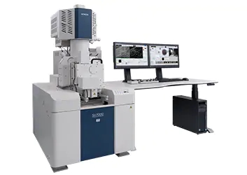
新型肖特基场发射扫描电镜SU7000
-可获得样品的各种信息,实现高通量分析-
肖特基场发射扫描电镜SU7000,它缩短了通过采集多种信号获取样品多种信息的时间,真正实现了高通量的观察与分析。

SU7000外观图
扫描电子显微镜(SEM)可通过检测样品激发出的二次电子、背散射电子、X射线等信号,获得从微细结构到组成成分等各种信息,因此被广泛应用于纳米技术、半导体、电子器件、生物、材料等诸多领域。随着SEM的应用范围在不断扩大,对观察时间的缩短、信号的迅速高效采集提出了更进一步的需求。
SU7000采用全新设计的探测器,使得对二次电子信号、背散射电子信号的检测以及分离能力大大提升。以前我们要根据获得的信号来调整样品与透镜之间的距离(工作距离/以下简称WD),以设置合适的观察与分析条件,而SU7000通过新研发的样品仓以及检测器系统,可在不改变WD的条件下更高效地接收各种信号,缩短了样品观察和分析的时间,提高了测试效率。
而且,SU7000还配置了可同时6通道显示界面(前代机型只能同时显示4通道),进一步升级SEM控制系统,大幅提高了信号获取速度,由此实现了样品的高通量观察。
它还标配超大样品仓,增设了附件接口,可适用于各种样品的观察与分析。
日立高新技术将在8月5日(星期日)~8月9日(星期四)在美国马里兰州举办的“Microscopy & Microanalysis”及9月5日(星期三)~9月7日(星期五)在幕张展示中心(千叶县千叶市)举办的“JASIS 2018”上展示这款SU7000,预计每年全球销量有望达150台。
【主要特点】
1.在相同WD的条件下,可同时实现二次电子、背散射电子观察与X射线分析
2.最多可同时实现6通道检测与显示
3.在高像素10,240 x 7,680时,也可获得图像数据
4.同级别*设备中最多的可配置18个附件接口
5.支持最低300Pa的低真空模式(选配)
*空间分辨率在1 nm/1 kV以下
【主要规格】
产品名称 | SU7000 |
电子源 | ZrO/W热场发射(肖特基热场发射) |
二次电子分辨率 | 0.8 nm(加速电压 15 kV) 0.9 nm(加速电压 1 kV) |
加速电压 | 0.1~30 kV |
放大倍率 | 20~2,000,000倍 |
束流 | 最大200 nA |
样品台 | X/Y/Z : 135 x 100 x 40 (mm) |
The advanced detection system of the SU7000 streamlines acquisition of structural, topographical, compositional, crystallographic, and other types of information by minimizing changes to microscope conditions, such as working distance or accelerating voltage.
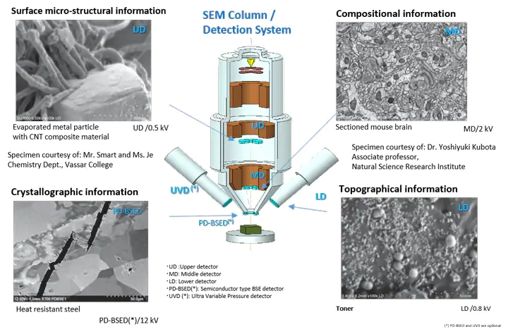
Specimen: Organic-coated gold rods
Specimen courtesy of: Mr. Smart and Ms. Je Chemistry Dept.,
Vassar College
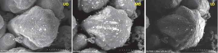
Simultaneous image acquisition for surface micro-structural information (UD), surface coating (MD), and overall topographic information (LD). Acceleration voltage: 1 kV
The software is capable of display 1, 2, or 4 signals including the chamber scope or SEM MAP on a single monitor.
Additionally, the operation panel can be customized to display submenus anywhere on the screen.
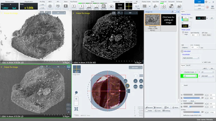
Dual Monitor
The first monitor can be used as a dedicated image display while the second monitor is utilized for operation.
Five detector images (UD, LD, UVD, MD, and PD-BSED) and SEM MAP of non-metallic inclusions in a steel specimen are displayed (left).
The screen shows the operation panel menu and the thumbnail image window on one screen (right).
The dual-monitor configuration supports increased productivity with expanded w
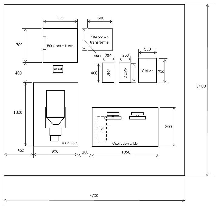
The specimen chamber can accommodate a tall specimen of φ 200 mm or 80 mm in height and 18 accessory ports. The large stage travels 135 mm (X) x 100 mm (Y) and can accept up to 2 kg of specimen.(*) Large specimen or variable type of sub-stages can be easily mounted on the front-opening large stage door.
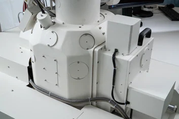
external view of the specimen chamber featuring 18 accessories por
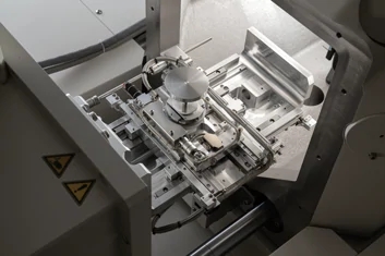
external view of the stage. XY movable range: 135×100 mm
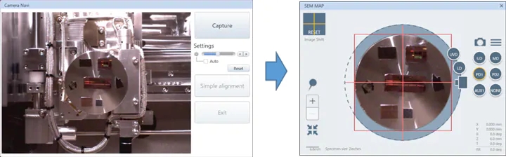
Left: Picture of the sample captured by the camera equipped inside the chamber.
Right: Camera image transferred to the SEM MAP screen for navigation.
The camera navigation feature correlates an optical image to the target observation area.
The camera installed in the specimen chamber captures the specimen image at the time of specimen introduction. The image is transferred to the SEM MAP screen for a graphically driven navigation interface.
Camera navigation supports a maximum of φ 100 mm specimen.
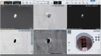
The SU7000 supports observation under various environmental conditions. A variety of detectors (*) such as UVD and MD are selectable in addition to the PD-BSED for observation under low-vacuum conditions.

Detector Selection Under Low-Vacuum Conditions
Specimen: Fiber with metallic oxide
Left: MD (Backscattered electron) image
Right: UVD (SE image)
The oxide dispersion and fiber layering state are observed respectively.
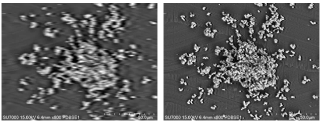
Improved PD-BSED Response Speed
Left: Traditional PD-BSED response at the scan rate of 30 ms x 64 frames
Right: SU7000 PD-BSED image demonstrating improved response and image quality to expand in-situ observation capability
| Image Resolution | Resolution SE | 0.8 nm@15 kV | |
|---|---|---|---|
| 0.9 nm@1 kV | |||
| Magnification | 20~2,000,000 x | ||
| Electron Optics | Emitter | ZrO/W Schottky Emitter | |
| Accelerating Voltage | 0.1~30 kV (0.01 kV step) | ||
| Probe Current | Max. 200 nA | ||
| Detectors | Standard Detectors | UD(Upper Detector) | |
| MD(Middle Detector) | |||
| LD(Lower Detector) | |||
| Optional Detectors | PD-BSED(Semiconductor type) | ||
| UVD (Ultra Variable Pressure Detector) | |||
| Variable Pressure(VP) Mode (Option) | Pressure Range | 5~300 Pa | |
| Available Detectors in VP mode | PD-BSED, UVD, UD, MD,LD | ||
| Specimen Stage | Stage Control | 5-axis Motor Drive | |
| Movable Range | X | 0~135 mm | |
| Y | 0~100 mm | ||
| Z | 1.5~40 mm | ||
| T | -5~70° | ||
| R | 360° | ||
| Specimen Chamber | Specimen Size | Max. φ200 mm, Max. 80mm Height | |
| Monitor(Option) | 23 inch LCD(1,920×1,080) , supports dual monitors operation | ||
| Image Display Mode | Large Screen Display Mode | 1,280×960 pixels | |
| Single Image Display Mode | 800×600 pixels | ||
| Dual Image Display Mode | 800×600 pixels、1,280×960 pixels with dual monitors | ||
| Quad Image Display Mode | 640×480 pixels | ||
| Hex Image Display Mode w/dual monitors | 640×480 pixels with dual monitors | ||
| Image Data Saving | Pixel Size | 640×480、1,280×960、2,560×1,920、5,120×3,840、10,240×7,680 | |
| Optional Accessories | Energy Dispersive X-ray Spectrometer (EDX) | ||
| Wavelength Dispersive X-ray Spectrometer (WDX) | |||
| Electron Backscattered Diffraction Detector (EBSD) | |||
| Cathodoluminescence System (CL) | |||
| Cryogenic Transfer System | |||
| Compatible with various types of sub-stages | |||